The Definition of Keratoconus
Keratoconus is a congenital disease of the cornea (autosomal dominant or autosomal recessive) and it belongs to the large group of hereditary corneal dystrophies.
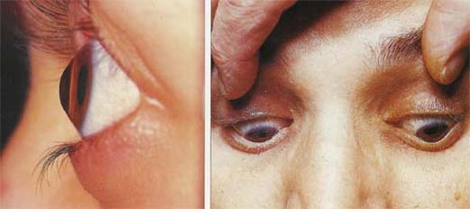
The disease is characterised by thinning and conuslike protrusion of the cornea. This is due to alterations that cause weakening of the corneal structure.
At first, the protrusion occurs in the inferior parts (lower half) but later it affects also the central part of the cornea.
Keratoconus mostly happens to be bilateral but often the progression is asymmetrical.
First symptoms generally start during puberty or early adolescence. In most cases patients realise a decrease in vision, or an increasing myopia with a significant progression of astigmatism can be found.
The incidence of keratoconus in the general population is about 1 in 2000.
The indications for Corneal Cross Linking
Keratoconus patients have to be checked / examined every 6-12 months.
If the findings are stable, treatment with glasses or contact lenses is sufficient.
If there is a progression an early cross linking treatment is recommended / indicated
This procedure stops the progression of the Keratoconus and therefore the need for penetrating keratoplasty (corneal transplant) could be significantly reduced in the future.
The treatment is relatively easy and low in costs.
Corneal Cross Linking
The aim of the treatment is to stabilise the cornea and prevent the progression of keratoconus.
The main structure of the corneal tissue (stroma) consists of single collagen fibers which are linked /inter connected.
The treatment of keratoconus with collagen cross linking is based on a significant stiffening of the corneal stroma due to photochemical cross linking of the single collagen fibers.
Therefore the single fibers form a "denser network" which leads to an increase in the overall stability of the cornea.
This procedure is performed under topical anaesthesia.
After an epithelium abrasion (corneal scraping), Riboflavin (vitamin B) drops are applied to the corneal surface over a period of 30 min and the cornea is then exposed to 365nm for 30min period thereafter.
During the follow up of eyes so far treated with collagen cross linking very few of the patients showed any more progression
In approximately 80% of the patients a regression of the maximal K-values (regression of the keratoconus) could be seen.
Post surgical corrected visual acuity improvement of 1 to 2 lines of Snellen lines can be expected.
No unwanted side effects such as opacification of the lens or loss of endothelial cells has been reported.
Only during the first 2 to 3 months after the cross linking a slight superficial corneal haze occurred.
Generally this minor haze disappears without any treatment, but a supportive therapy with soothing ointment can be used.